The convenience of snapping a photo and reading a digital chart on a smartphone screen has radically transformed how Canadians renew their contact lens prescriptions, especially as the brisk autumn air settles in and schedules tighten. For one Toronto graphic designer, avoiding the typical clinic wait time and the 20-kilometre commute seemed like the ultimate modern life hack. After breezing through a popular mobile application to quickly update his astigmatism script, he received an automated, algorithmically generated ‘green light’ to purchase his yearly supply. But beneath the surface of that seemingly flawless, frictionless checkout process, a silent, painless threat was rapidly expanding in the deep shadows of his peripheral vision, entirely undetected by the camera lens of his device.
Just three days after his digital renewal cleared, a fleeting shadow—described as an eerie, dark curtain slowly creeping over the outer edge of his visual field—prompted a panicked, emergency walk-in visit to a local clinical centre. It was here that a seasoned medical specialist caught exactly what the software algorithm had completely missed: a massive, horseshoe-shaped tear in the neurosensory tissue that was literally hours away from causing permanent, irreversible blindness. This terrifying clinical crisis has sparked a massive, urgent warning from Ontario Optometrists, who are currently reporting a drastic, unprecedented surge in silent, sight-threatening conditions passing seamlessly through the cracks of these automated digital platforms.
The Hidden Dangers of Algorithmic Eye Care
As temperatures frequently drop below zero Celsius across the country, the allure of staying indoors and using an application to handle health logistics is undeniably strong. Marketing campaigns for online vision testing platforms heavily emphasize speed, convenience, and cost-reduction, aggressively targeting busy professionals and university students. However, studies confirm that these platforms fundamentally confuse ‘visual acuity’—the simple mechanical ability to read high-contrast black letters against a stark white background lacking any subtle colour contrast—with comprehensive ocular health. A simple camera flash simply cannot illuminate the complex, multi-layered physiological universe that exists at the back of the human eye.
When Ontario Optometrists evaluate a patient in person, they are not merely checking if a localized optical prescription requires a minor adjustment of a few diopters. They are actively screening for systemic vascular diseases, microscopic inflammatory markers, and early-stage neurological anomalies that manifest first within the ocular microcirculation. Automated systems lack the stereoscopic depth perception and the high-magnification optical coherence necessary to detect structural abnormalities that do not immediately alter central vision. This creates a dangerous false sense of security for the end-user relying entirely on algorithmic assumptions.
Comparing the Care Modalities
| Care Modality | Target Audience | Marketed Benefits | Hidden Clinical Risks |
|---|---|---|---|
| Automated Online Vision Apps | Busy adults, students, routine contact lens wearers | Zero travel time, instant prescription renewals, perceived lower cost | Zero evaluation of internal retinal health, misses asymptomatic tears, ignores corneal oxygen deprivation |
| In-Person Clinical Examination | All demographics, especially those with high myopia or systemic health issues | Comprehensive health baseline, precise astigmatism mapping, immediate crisis intervention | Requires scheduling, potential wait times, commuting logistics |
To truly grasp how these digital platforms fail the strictly regulated medical grade, one must look deep into the microscopic anatomy of our most vital sensory organ and the hidden mechanics of internal optical failure.
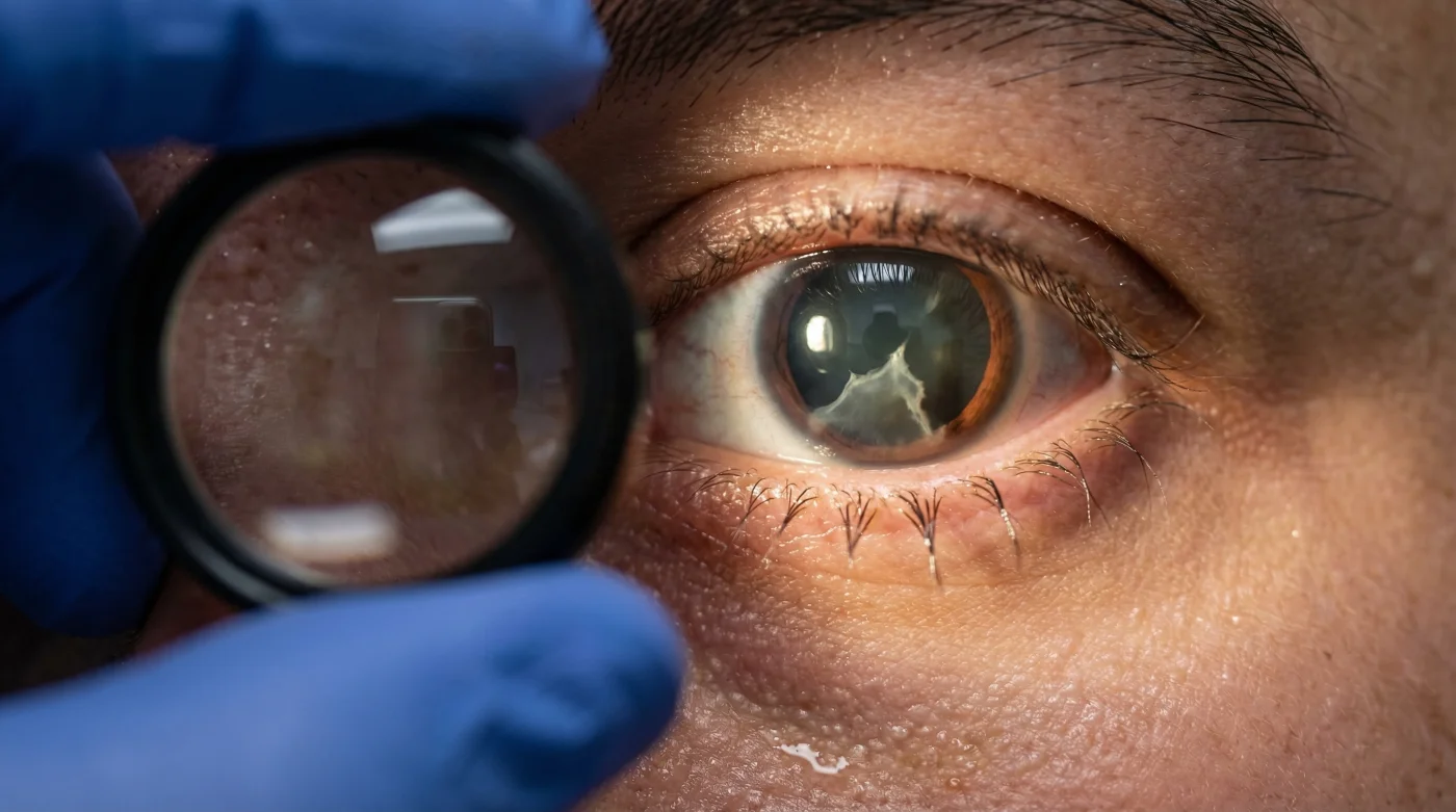
The Anatomy of a Missed Retinal Tear
The interior of the human eye is filled with a dense, gel-like substance known as the corpus vitreum, or vitreous humour. As we age, or in individuals who are highly nearsighted, this gel undergoes a natural, unpredictable liquefaction process. Over time, the vitreous can begin to shrink and pull away from the delicate, light-sensitive wallpaper lining the back of the eye—the retina. While this separation is common, an abnormally strong adhesion can cause the shrinking gel to physically rip the neurosensory tissue, creating a retinal tear. Because the retina completely lacks pain receptors, this catastrophic structural failure occurs in total silence, generating no physical discomfort whatsoever.
This is precisely why algorithms evaluating central reading ability fail so spectacularly. A tear usually originates in the far retinal periphery. A patient can easily read the standard 20/20 line on a glowing smartphone screen while a massive tear allows liquefied vitreous to seep underneath the tissue, slowly peeling it away like wet wallpaper. Experts advise that relying solely on central acuity checks is fundamentally akin to verifying a car’s tire pressure while the engine is actively catching fire.
Diagnostic Troubleshooting: Symptom = Cause
- Ayahuasca ceremony resulted in Connor Murphy’s total career pivot
- Blood Moon 2026 turns the sky red over Canada tomorrow morning
- Ceramic Shield 2 glass on iPhone 17e resists three times more scratches?
- British Columbia residents will see the entire Blood Moon tonight
- Connor Murphy shocked with his appearance using only sunlight and water
- Symptom: Sudden, explosive onset of translucent, web-like floaters. = Cause: Posterior Vitreous Detachment (PVD) causing microscopic cellular debris and condensed collagen fibres to cast internal shadows on the macular centre.
- Symptom: Brief, lightning-like flashes of light in the peripheral vision, especially noticeable in dark environments. = Cause: Physical, mechanical traction of the shrinking vitreous gel violently pulling against the neurosensory retinal layers, which the brain misinterprets as sudden bursts of light.
- Symptom: A heavy, opaque dark curtain or persistent shadow obscuring a portion of the visual field. = Cause: Amotio retinae (full retinal detachment), indicating that liquid has aggressively seeped under the initial tear, functionally blinding the affected photoreceptors.
The Clinical Data and Dosing Metrics
| Clinical Tool / Medication | Standard Dosing / Clinical Metric | Scientific Diagnostic Mechanism |
|---|---|---|
| Tropicamide (Mydriatic Agent) | 1.0% concentration drop; 20-30 minute incubation period | Temporarily paralyzes the iris sphincter muscle, dilating the pupil to 7-8 millimetres for panoramic peripheral viewing. |
| Optical Coherence Tomography (OCT) | Scans utilizing near-infrared light (840 nm wavelength); measuring tissue down to 5 microns | Generates highly detailed, cross-sectional topographical maps of the macula and optic nerve head. |
| Goldmann Applanation Tonometry | Target pressure strictly remaining under 21 mmHg | Accurately measures intraocular pressure by calculating the exact physical force required to flatten a specific corneal area. |
Recognizing these internal biological alarm bells is absolutely critical, but securing a physical evaluation that actively deploys these precise diagnostic metrics forms the true foundation of long-term optical preservation.
Standardizing the Comprehensive Ocular Examination
When Ontario Optometrists actively advocate for routine, in-person physical examinations, they are demanding strict adherence to a standardized, rigorous clinical protocol that absolutely no piece of consumer software or mobile application can replicate. A truly comprehensive evaluation moves far beyond simply asking a patient to cover one eye and read a localized digital chart. It involves a systematic, multi-tiered clinical investigation into the structural and neurological integrity of the entire ocular cavity.
During a certified medical exam, the clinical environment is heavily controlled. Specific lighting is optimized to gauge subtle pupillary reflexes, and specialized magnifying equipment is brought in to thoroughly examine the anterior segment of the eye, ensuring that prolonged contact lens wear hasn’t triggered microscopic vascularization or silent corneal suffocation. For the graphic designer who nearly lost his vision, it was the immediate, urgent application of specialized lenses and intense clinical lighting that illuminated the deeply hidden crisis before it was too late.
The Top 3 Essential Diagnostic Steps
A premium clinical examination relies heavily on an unyielding hierarchy of procedural checks:
- The Slit-Lamp Biomicroscopy: A high-intensity light source coupled directly with a binocular microscope allows the practitioner to view the cornea, iris, and crystalline lens in extreme three-dimensional detail, diligently checking for early cataracts or inflammatory cells.
- Fundus Photography and Topography: Capturing high-resolution, ultra-widefield imagery of the entire retinal landscape to document baseline vascular patterns and track subtle, year-over-year microscopic changes.
- Pharmacological Dilation: Utilizing highly specific pharmaceutical drops to forcibly widen the pupil, effectively opening the anatomical ‘door’ to the interior chamber so that the far peripheral edges of the retina can be physically inspected with a stereoscopic headset.
The Optical Quality Guide
| Practice Component | What To Look For (Green Flags) | What To Avoid (Red Flags) |
|---|---|---|
| Diagnostic Equipment | Presence of ultra-widefield imaging, 3D OCT scanning capabilities, and modern applanation tonometry. | Practices relying solely on outdated, automated ‘puff-of-air’ pressure checks and basic wall charts. |
| Practitioner Interaction | Detailed history taking, thorough discussions about systemic health (diabetes, hypertension), and transparent explanation of microscopic imaging. | Rushed, highly transactional five-minute encounters focused entirely on finalizing a lens prescription for a quick retail sale. |
| Prescription Transparency | Provision of a comprehensive medical document detailing base curve, diameter, and optimal lens material for critical oxygen breathability. | Automated digital approvals that bypass physical corneal fit and health assessments entirely. |
Arming yourself with this deep clinical knowledge is only the first phase; executing a strict, preventative routine is what ultimately shields and saves your biological sight.
A Preventative Plan for Long-Term Vision Preservation
The highly marketed convenience of modern technology should always serve as a helpful supplement to our daily lives, not a dangerous, corner-cutting replacement for specialized medical care. The stark, terrifying reality faced by the Toronto designer serves as a highly visible, critical warning to all Canadians currently relying on automated mobile algorithms for their essential biological well-being. Studies confirm that rapid, early intervention in cases of asymptomatic retinal tearing yields a clinical success rate of well over 95%, typically involving a quick, localized, outpatient laser procedure known as laser retinopexy, which precisely welds the torn tissue back into its proper anatomical position. However, this high success rate drops exponentially with every single passing hour the condition remains undiagnosed.
Experts advise scheduling a comprehensive, fully dilated eye examination every twelve to twenty-four months, specifically depending on your individual systemic risk factors, age, and natural refractive error. If you happen to experience any sudden onset of the diagnostic symptoms outlined above—specifically the dark curtains, floating translucent debris, or peripheral lightning flashes—you must immediately bypass all digital conveniences and seek urgent, emergency physical care. Ontario Optometrists continue to stand firmly at the frontline of localized optical health, deeply armed with the scientific metrics, rigorous clinical standards, and the sophisticated diagnostic technology necessary to heavily protect your eyes from the unseen, silent threats currently lurking in the biological shadows.
By intentionally prioritizing rigorous medical evaluation over fleeting digital shortcuts, you successfully build an unbreakable bridge between today’s clinical caution and a lifetime of vibrant, uncompromised vision.
Read More